
LAMENESS INVESTIGATIONS
There are 3 stages to a lameness investigation:
- Where is the lameness coming from?
- What is the cause of the lameness?
- What can we do about the lameness?
Stage 1: Where is the lameness coming from?
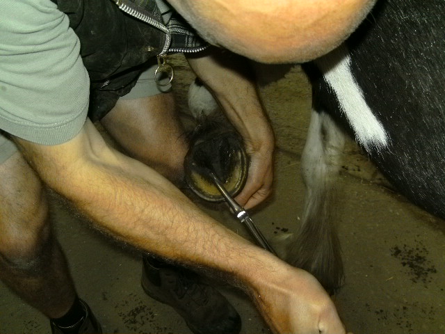
Nick uses hoof testers to find any sore areas on a foot
This is often the most difficult stage. The process would be much quicker if you could just ask the horse where it hurts! Often an initial physical exam will reveal areas of swelling, heat or pain which indicate certain areas. However, it is easy to be caught out and the basic principle at this stage is to make the horse sound – whatever region you have made pain free is then the source of pain. This is done by nerve or joint blocks with local anaesthetic.
Nerve blocks: local anaesthetic is deposited around a nerve. If the nerve supplies the painful area then the horse will go sound. This sounds very straightforward but often it is not. In thick-skinned cobs with thick feathers, the nerve can be hard to find. If the lameness is improved but not completely alleviated, are there two problems or did the block not work very well? Experience in interpretation is essential.
Joint blocks: local anaesthetic is injected directly into a suspected joint. There are risks associated with this technique and in the worst case scenario a joint infection could result. However, every effort is made to make the injection sterile and this is a routine procedure. Even here there are difficulties with interpretation. Uncomplicated joint pain should block out in 5-7 minutes. Sometimes it takes longer and the significance of this must be assessed.
The standard procedure in a lameness investigation is to start at the bottom and work up. However, this can result in a long, drawn-out investigation if the lameness is in the stifle for example. Often we will take a considered short cut in the interests of cost effectiveness. The most common example of this is the hocks. If a middle aged horse has hind-limb lameness and is positive to a flexion test then we may decide to go straight to a hock X-rays.
Occasionally, the blocks will reveal nothing useful. In these cases it is possible that muscle pain is the source of the pain (although this would hopefully be picked up by the physical examination).
In refractory cases it may be worth using scintigraphy (a ‘bone scan’). In this technique, the horse is injected with a mildly radioactive element which is actively taken up by bone. Traumatized bone is very active and takes up more. When a gamma camera is used the whole horse can be viewed and any radioactivity hotspots will indicate an area of special interest. Scintigraphy does not generally give you a diagnosis but it does tell you where to focus on.
What is the cause of the lameness?
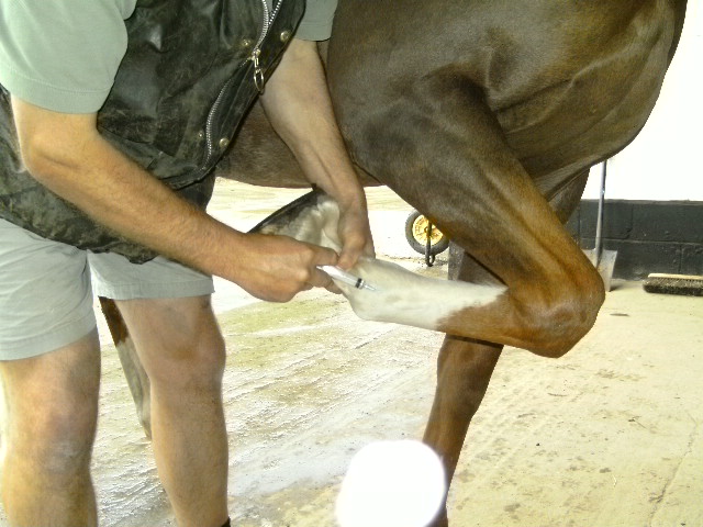
Nick performs a simple nerve block
Once we have found the anatomical site of the pain then the next step is to work out what is causing the pain. Radiography and ultrasound are still the main imaging techniques. For example, a lameness that blocks to the fetlock would then be x-rayed. However, lameness that blocks to a tendon sheath would require an ultrasound evaluation since soft tissues are more likely to be involved. Often, both techniques would be used. MRI is now available for lower leg problems as a referral procedure.
What can we do about it?
Naturally this is the part you are most interested in. Once we know ‘where and why’, we then have to decide what we can do about it and what is the horse’s prognosis for future performance. Treatment may be as simple as rest, anti-inflammatories and then a controlled return to work. Direct medication of a problem joint with steroids may be indicated. Arthroscopy may be the best course of action. Here, a camera is put into the joint and the problem can be directly visualized. Arthroscopy is often a final diagnosis and a treatment; when the surgeon is in the joint, loose bits of cartilage can be removed for example.